apical lung hernia
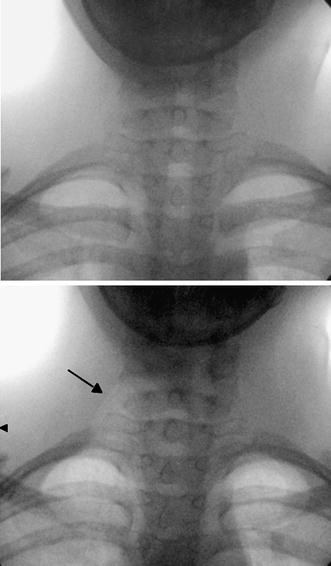
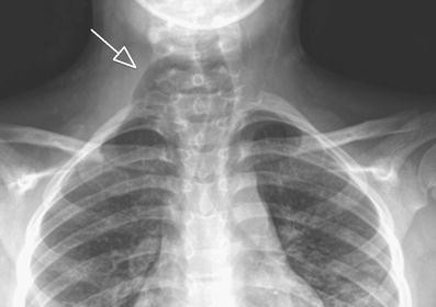
Apical lung hernias typically manifest as unilateral right-sided air radiolucencies at the thoracic inlet on chest radiographs and can cause lateral tracheal deviation. About half of all lung hernias however appear after trauma to the chest 5.

Article Medicale Tunisie Article Medicale
To the Editor.

. 1 A hernia involving the cervical space has also been described as apical in the literature. CONCLUSION Apical lung hernias typically manifest as unilateral right-sided air radiolucencies. Apical Lung Hernia.
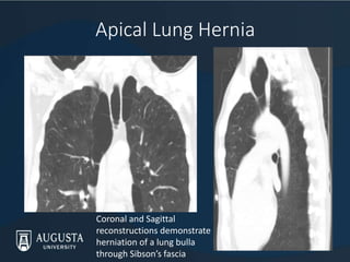
Apical lung hernia is a rare variety and has been confined to few case reports and series. Spontaneous apical lung herniation presenting as a neck lump in a patient with Ehlers-Danlos syndrome Ehlers-Danlos syndrome EDS is a heritable group of disorders of connective tissue characterised by skin hyperlaxity joint hypermobility and tissue fragility. Although lung hernias usually do not cause problems knowledge of their presentation on either neck or chest radiographs or CT Fig.
Airway fluoroscopy or CT pe. Vided the stimulus to develop a new treatment of direct Chest 199098987 8. Pulmonary hernias have been described through the diaphragm intercostal spaces and into the cervical space.
Institutional and Research4Life access to the MJA is now provided through Wiley Online Library. Sometimes the diagnosis can only be made with a Valsalva maneuver which accentuates the herniation improving its visibility on physical examination. Dysphagia esophageal or coughing trachea 2.
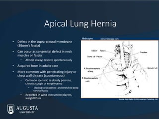
DISCUSSION Apical lung herniation in adults is rare particularly in the absence of penetrating lung injury or chest wall disease1 2 It is due to a defect in the suprapleural membrane Sibsons fascia and small incidental apical parietal pleural defects have been. Some hernias of the lung however are symptomatic being accompanied by local pain paroxysmal coughing hemoptysis or any combination of the three. The term pathological hernia is used to describe lung herniation induced by conditions such as tuberculosis focal bacterial infections empyema or osteomyelitis and malignant diseases 1.
17874988 Indexed for MEDLINE Publication Types. Developed by renowned radiologists in each specialty STATdx provides comprehensive decision support you can rely on - Apical Lung Hernia. Apical lung hernias typically manifest as unilateral right-sided air radiolucencies at the thoracic inlet on chest radiographs.
Symptoms when reported tend to be due to extrinsic pressure from the hernia on neck structures eg. Am J Radiol 199616792730. Showed an apical lung hernia at the right apex figure 2AeC.
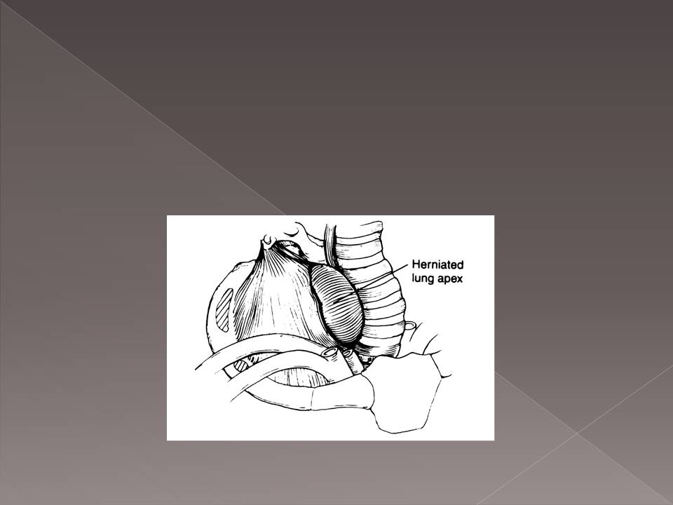
They are frequently intermittent and can cause lateral tracheal deviation. Dysphagia esophageal or coughing trachea 2. Herniation occurs through a defect in the Sibsons fascia and the apical segment of the lung protrudes in between the scalenus anterior and sternocleidomastoid muscles.
We performed this study to characterize the clinical and radiologic manifestations of apical lung hernias. Symptoms when reported tend to be due to extrinsic pressure from the hernia on neck structures eg. OBJECTIVE We performed this study to characterize the clinical and radiologic manifestations of apical lung hernias.
Hernia of the lung occurs infrequently and not all of those that do occur cause symptoms that require treatment. Air in the neck. Apical lung hernias are often asymptomatic 1-3.
Radiologic studies performed at midinspiration may not show hernias. Apical lung herniation in adults is rare particularly in the absence of penetrating lung injury or chest wall disease1 2 It is due to a defect in the suprapleural membrane Sibsons fascia and small incidental apical parietal pleural defects have been described which may be present prior to the development of a larger defect3 4 Tearing of the fascia and spontaneous. Radiological findings in six cases.
4-Chest radiograph shows bilateral congenital apical lung hernia- scans may avoid complications that may arise from insertion tion greater on. Apical lung hernias are often asymptomatic 1-3. Laryngocele apical lung hernia and apical paraseptal blebsor bullae Lung herniation is rare and can be described as the protrusion ofpulmonary tissueoutsidethethoraciccav.
They are frequently intermittent and can cause lateral tracheal deviation. In rare instances a lung hernia may become strangulated. The patient was discharged on the 9thpostoperative day in a good condition.
The full article is accessible to AMA members and paid subscribers. Sometimes the diagnosis can only be made with a Valsalva maneuver which accentuates the herniation improving its visibility on physical examination. Direct intraoperative nitrous oxide cryosurgery 4 pro- 6.
Apical lung hernia. Apical lung hernias typically manifest as unilateral right-sided air radi- olucencies at the thoracic inlet on chest radiographs. Control examination 2 years after the operation found no deformation of the thorax or recurrence of the lung hernia.
1Respiratory Medicine BYL Nair Hospital and Topiwala National Medical College Mumbai Maharashtra India. My attention was drawn to the Snapshot of an apical lung hernia published in the Journal last year. Li C Miller WT.
Login to read more or purchase a subscription now. They are frequently intermittent and. Roentgenogram of the month.
Pulmonary herniation is an extension of the lung and pleura beyond their native positions in the thoracic cavity. Apical lung hernias typically manifest as unilateral right-sided air radiolucencies at the thoracic inlet on chest radiographs. Conventional approach by left mid-lateral thoracotomy was used for surgical repair of the lung hernia and stabilization of the intercostal space.
Medical College of Georgia. Radiologic studies performed at midinspiration may not.

Apical Lung Herniation Radiology Case Radiopaedia Org

Cardiac Noninvasive Imaging Chest Radiography Cardiovascular Magnetic Resonance And Computed Tomography Of The Heart Springerlink
Epos Trade
Interesting Cases Of Lung Hernias

Chest X Ray Showed Apical Mass On The Right Lung No Pleural Effusion Download Scientific Diagram

Frontiers The Chest Radiographic Thoracic Area Can Serve As A Prediction Marker For Morbidity And Mortality In Infants With Congenital Diaphragmatic Hernia

Pdf Dynamic Cervical Lung Lobe Herniation In A Shih Tzu Dog Semantic Scholar

Massive Hiatal Hernia Of Gastrointestinal Tract A Rare Intrathoracic Gastrointestinal Disease Cheung 2009 Journal Of The American Geriatrics Society Wiley Online Library

Right Upper Lobe Pneumonia Pneumonia Radiology Medicine
Dynamic Cervical Lung Herniation In A 10 Year Old Girl With Cough Springerlink

Bilateral Cervical Lung Hernia With T1 Nerve Compression The Annals Of Thoracic Surgery

45 Year Old Asymptomatic Man With Right Apical Lung Hernia Download Scientific Diagram
![]()
Extra Luminal Contrast Leakage Was Detected On The Initial Esophagogram Download Scientific Diagram
Dynamic Cervical Lung Herniation In A 10 Year Old Girl With Cough Springerlink

Is Collapse Of The Lung With Increased Lucency On A Chest X Ray Always A Pneumothorax Journal Of Cardiothoracic And Vascular Anesthesia

Right Sided Congestive Heart Failure Secondary To Supraventricular Tachycardia In A Dog With A Right Atrial Mass Basili 2021 Veterinary Record Case Reports Wiley Online Library
Interesting Cases Of Lung Hernias

Intercostal Lung Hernia Radiographic And Mdct Findings Clinical Radiology
Pdf Adult Cervical Lung Herniation Importance Of Valsalva Manoeuvre In Imaging Semantic Scholar


Comments
Post a Comment